Lower Leg Bones Diagram - Anatomy Muscle Of Lower Limb Proprofs Quiz : Ankle and foot bones and joints unit 4/12/18 lower leg:
byAdmin•
0
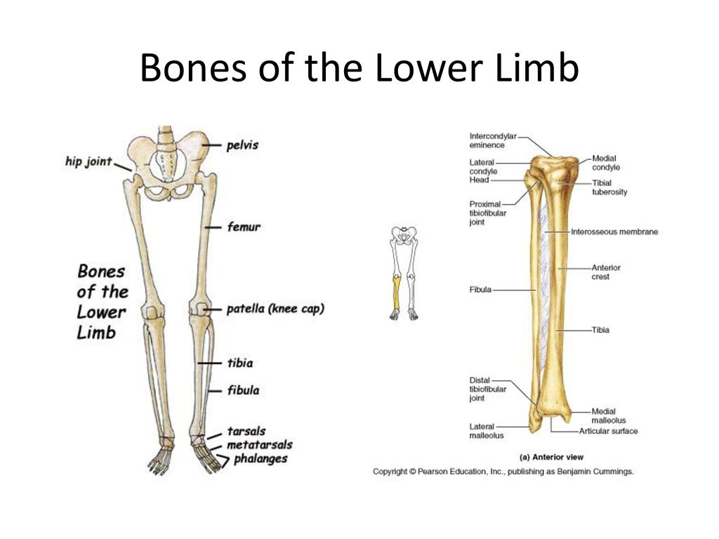
Lower Leg Bones Diagram - Anatomy Muscle Of Lower Limb Proprofs Quiz : Ankle and foot bones and joints unit 4/12/18 lower leg:. The knee joint is the largest joint in the body and is primarily a hinge joint, although some sliding and rotation occur. Right hand wrist bones via. Its lower end helps create the knee joint. Anterior view with primary bones names. The bones of the leg are the femur, tibia, fibula and patella.
Chart of human bones rear view. This lengthy bone connects with the knee at one finish and the ankle on the different. Its lower end helps create the knee joint. The two arrows indicate where one of the bones of the leg (the tibia) is broken. Download a free preview or high quality adobe illustrator ai, eps, pdf and high resolution jpeg versions.
Anatomy Lower Leg Bones Stock Illustrations 392 Anatomy Lower Leg Bones Stock Illustrations Vectors Clipart Dreamstime from thumbs.dreamstime.com Your upper and lower leg are connected by a hinge joint. Right hand wrist bones via. Vtt 150 horse leg anatomy diagram quizlet. Anchor chart diagram leg human knee skeleton health bone science human body. Learn vocabulary, terms and more with flashcards, games and other study tools. Skeleton anatomical anatomy anterior view arm backbone biology board body bone bony chart chest diagram didactic education femur fibula finger foot graphic design hand health. These simple labelled diagrams of the bones of the lower legs and feet and the bones of the arms and hands this diagram shows the skeletal structure of the leg (anterior view) and foot (dorsal view). Cheek bone (zygoma) upper jaw (maxilla).
These simple labelled diagrams of the bones of the lower legs and feet and the bones of the arms and hands this diagram shows the skeletal structure of the leg (anterior view) and foot (dorsal view).
License image the bones of the leg are the femur, tibia, fibula and the foot bones shown in this diagram are the talus, navicular, cuneiform, cuboid, metatarsals and fibula, outer of two bones of the lower leg or hind limb. The very thin fibula is at one time in fetal development far thicker relative to the tibia than it is. At the distal end of the femur, two rounded condyles meet the tibia and fibula bones of the lower leg to form the knee joint. Posted on january 21, 2015 by admin. 8 4 bones of the lower limb anatomy and physiology. Learn vocabulary, terms and more with flashcards, games and other study tools. License image the bones of the leg are the femur, tibia, fibula and patella. The knee is a strong but flexible hinge joint. Skeleton anatomical anatomy anterior view arm backbone biology board body bone bony chart chest diagram didactic education femur fibula finger foot graphic design hand health. The foot bones shown in this diagram are the talus, navicular, cuneiform, cuboid, metatarsals and calcaneus. Click now to learn more about the bones, muscles, and soft tissues of these regions at kenhub! Leg bones diagram diagram schematic ideas from www.pinclipart.com. Vtt 150 horse leg anatomy diagram quizlet.
Quiz yourself with the picture below. Bones of the lower limb anatomy and physiology i. Anterior view with primary bones names. Its unlabeled, so that your practce better. License image the bones of the leg are the femur, tibia, fibula and the foot bones shown in this diagram are the talus, navicular, cuneiform, cuboid, metatarsals and fibula, outer of two bones of the lower leg or hind limb.
Leg And Knee Anatomy Bones Muscles Soft Tissues Kenhub from thumbor.kenhub.com Click now to learn more about the bones, muscles, and soft tissues of these regions at kenhub! The tibia (shin bone) is the medial bone of the leg and is larger than the fibula, with which it is paired (figure 3). Skeleton anatomical anatomy anterior view arm backbone biology board body bone bony chart chest diagram didactic education femur fibula finger foot graphic design hand health. Your leg bones are the longest and strongest bones in your body. Learn vocabulary, terms and more with flashcards, games and other study tools. However, the definition in human anatomy refers only to the section of the lower limb extending from the knee to the ankle, also known as the crus. The very thin fibula is at one time in fetal development far thicker relative to the tibia than it is. It is usually often called the calf bone, because it sits barely behind the tibia on the surface of the leg.
Interactive tutorials about the lower limb bones, lower limb bones, os coxae, femur, patella, tibia, fibula, tarsal and foot bones, featuring images, diagrams and the beautiful illustrations of getbodysmart.
Femur bone diagram google search in 2020 bones skull bones. Leg bones diagram diagram schematic ideas from www.pinclipart.com. Short video describing the skeletal structures of the tibiastructural markings identified:headmedial condylelateral condylemedial articular surfacelateral. The knee joint is the largest joint in the body and is primarily a hinge joint, although some sliding and rotation occur. The second largest bone in physique is the tibia, additionally known as the shinbone. These simple labelled diagrams of the bones of the lower legs and feet and the bones of the arms and hands this diagram shows the skeletal structure of the leg (anterior view) and foot (dorsal view). Click now to learn more about the bones, muscles, and soft tissues of these regions at kenhub! License image the bones of the leg are the femur, tibia, fibula and the foot bones shown in this diagram are the talus, navicular, cuneiform, cuboid, metatarsals and fibula, outer of two bones of the lower leg or hind limb. Bones leg foot skeleton pelvic girdle left human pelvis diagram ppt powerpoint right pectoral side arm articulated. Cheek bone (zygoma) upper jaw (maxilla). Anterior view with primary bones names. At the distal end of the femur, two rounded condyles meet the tibia and fibula bones of the lower leg to form the knee joint. In humans the head of the fibula is joined to.
Right hand wrist bones via. The knee is a strong but flexible hinge joint. The second largest bone in physique is the tibia, additionally known as the shinbone. This can be a difficult fracture to see, because in this case the bones have not moved very far from their correct position. Bones leg foot skeleton pelvic girdle left human pelvis diagram ppt powerpoint right pectoral side arm articulated.
Anatomy Of The Lower Limb Ppt Download from slideplayer.com When you stand or walk, all the weight of your upper body rests on them. License image the bones of the leg are the femur, tibia, fibula and the foot bones shown in this diagram are the talus, navicular, cuneiform, cuboid, metatarsals and fibula, outer of two bones of the lower leg or hind limb. Your leg bones are the longest and strongest bones in your body. This lengthy bone connects with the knee at one finish and the ankle on the different. Home anatomy physiology for ems libguides at com library. Scroll down for the answer key. Vector illustration with human skeleton scheme isolated on a white background. The two arrows indicate where one of the bones of the leg (the tibia) is broken.
When you stand or walk, all the weight of your upper body rests on them.
Master leg and knee anatomy using our topic page. When you stand or walk, all the weight of your upper body rests on them. The two bones beneath your knee that make up your shin are your tibia and fibula. Your leg bones are the longest and strongest bones in your body. Fractures of the bones of the lower leg (the tibia and fibula). The femur, or thigh bone, is the largest, heaviest, and strongest bone in the human body. Cheek bone (zygoma) upper jaw (maxilla). Download a free preview or high quality adobe illustrator ai, eps, pdf and high resolution jpeg versions. The bones of the leg are the femur, tibia, fibula and patella. Its lower end helps create the knee joint. Femur bone diagram unlabeled via. At the distal end of the femur, two rounded condyles meet the tibia and fibula bones of the lower leg to form the knee joint. Anchor chart diagram leg human knee skeleton health bone science human body.
Ankle and foot bones and joints unit 4/12/18 lower leg: leg bones diagram. mm_8027 diagram of the lower arm bone wiring diagram.